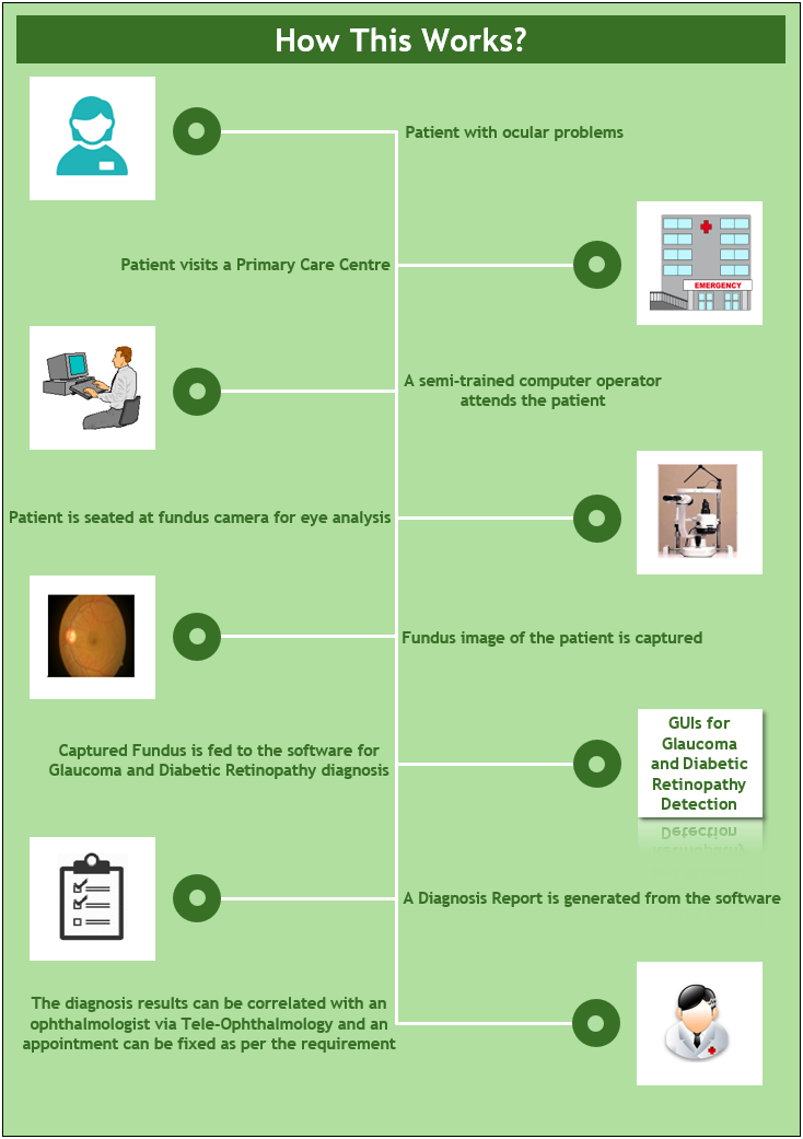
A person having some problems in his/her eyesight may visit a primary care center or eye hospital. The person is attended by a technical operator and asks the patient to sit in front of a high-resolution fundus camera for examination. The operator will focus the camera and capture the fundus image. Once, the image is taken, it is directly fed to the developed softwares. Some basic information like name, gender and age are fed to the software for future references. A diagnostic test is performed for detection of Glaucoma or Diabetic Retinopathy on the fundus image. Instantly, a diagnosis report is generated with detailed information of clinical parameters determined using the image processing techniques. If any signs of disease are present in the report, then a correlation of the same can be done by a trained ophthalmologist, who is available at a remote location. And depending on ophthalmologist’s views, an appointment can be fixed, if required.