FOLLOW

About

Human Eye
FUNDUS IMAGE
GLAUCOMA
What is Glaucoma?

Glaucoma is an optic neuropathy which can cause damage to the optic nerve and result in permanent vision loss.

Under normal scenarios, the circulation cycle of aqueous humor is performed well. However, when some aberrations occur, this fluid is not able to flow out. This causes a blockage in the channel and causes a pressure inside the eye. This intra-ocular pressure creates an inward force on the retina.

Usually, an optic disc is a flat structure, which can become non-flat due to increased IOP. The pressure causes the disc to bend and results in cupping of the disc. This is clinically termed as Optic Cup.

Challenges in detection of Glaucoma using Image Processing

Why Image Processing?

Currently, a clinical diagnosis for Glaucoma detection is performed under the supervision of a trained ophthalmologist. There is no such tool designed yet which can give a decision in absence of medical expert. So, an automated diagnostic tool can be used for screening purpose at primary care centres. Such a tool can act as an assisting tool and screen the affected patients so that proper medical care can be provided to them.

The medical signs such as cupping of optic disc, haemorrhages, blood vessel abnormality, peri-papillary atrophy and RNFL defects are some clinical observations used by medical experts across the globe. These clinical observations impart visual changes to a fundus image and can easily be differentiated from an image which has no signs.

Visual changes like change in intensity and colour of pixels, tortuosity of blood vessels can be observed which can be easily detected using IMAGE PROCESSING techniques. Also, the diagnosis is done by considering the clinical input from medical experts, so there are less chances of wrong diagnosis.

Challenges

Glaucoma is a disease majorly related to the optic nerve head region. Structural changes may occur in the optic nerve head region which may characterize the presence of Glaucoma. Since, the disease is related to optic nerve head region, so this region should be segmented accurately using image processing techniques. However, the presence of medical signs like bright lesions (exudates and cotton soft wools) and some artefacts which gets introduced in the images during acquisition process, affect the accuracy of optic disc segmentation. The following can be considered some important challenges which are faced during ONH segmentation:

  • Exudates
  • Reflections (artefacts introduced due to illumination)
  • Correct ONH localization
  • Choroid Vessels
  • Blood Vessels overlaying on the ONH region
Block Diagram for Glaucoma detection:
Highlights
Correct ONH Localization from non-uniformly illuminated images
Correct Rejection of False Positives
Blood Vessel Inpainting for accurate optic cup boundary detection
Adaptive Intensity based threshold based on local features from image
Instant generation of Diagnosis Report with information about clinical parameters
Grade an image into 3 classes to provide medical care accordingly
Fix appointment with an Ophthalmologist if test appears positive for Glaucoma
Clinical Parameters used for Glaucoma Detection

The use of 3 different clinical parameters make SOP-G robust in giving a decision for a fundus image.

Area Cup-to-disc ratio:


The expansion of optic cup is determined by expressing the cup area in terms of disc area

Vertical Cup-to-disc ratio:


The expansion of optic cup is determined along the vertical direction

ISNT Rule:


A detailed analysis of the structural changes in ONH due to Glaucoma

Grading of a Fundus Image

Post diagnosis, a report is generated with a decision regarding the class of the tested fundus image. With the use of the 3 clinical parameters, the fundus images are classified as follows:

NORMAL:


The clinical parameters are within a specified range. The patient does not have to worry.

SUSPECT GLAUCOMA:


The patient has shown signs of Glaucoma and progression is in early stage.

GLAUCOMA:


The disease has progressed in an advanced stage. Proper medical care is needed.
Diagnosis Report:

PATIENT DETAILS:


  • Name (Characters only)
  • Age (Numbers only)
  • Gender (Male / Female)

IDENTIFIER DETAILS:


  • Date (System generated)
  • Time (System generated)
  • Patient ID

PARAMETER DETAILS:


  • Area CDR
  • Vertical CDR
  • NRR Area in ISNT Quadrants

NOTE: The diagnosis report should be clinically correlated with an Ophthalmologist.

DIABETIC RETINOPATHY
Introduction of Diabetic Retinopathy

Diabetic retinopathy also known as diabetic eye disease, is a medical condition in which damage occurs to the retina due to diabetes. It can eventually lead to vision loss and blindness.

  • Progressive dysfunction of the retinal blood vessels caused by chronic hyperglycemia
  • DR can be a complication of diabetes type 1 or diabetes type 2
  • Initially, DR is asymptomatic, if not treated though it can cause low vision and blindness.
  • Diabetic retinopathy is responsible for 1.8 million of the 37 million cases of blindness throughout the world
  • People with diabetes are 25 times more likely to become blind than the general population.
Changes observed in Eye affected from DR.
  • All forms of diabetic eye disease have the potential to cause severe vision loss and blindness.
  • Diabetic retinopathy involves changes to retinal blood vessels that can cause them to bleed or leak fluid, distorting vision.

Fundus image with DR abnormalities

Stages of Diabetic Retinopathy
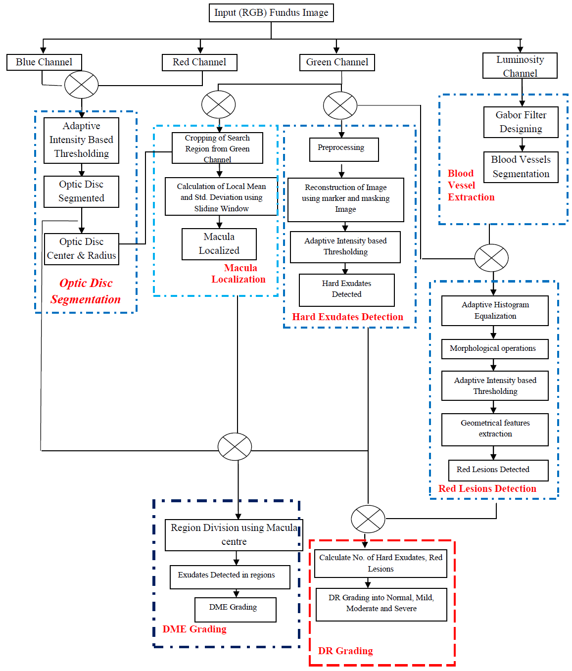
Block Diagram for Diabetic Retinopathy Detection
Challenges in detection of DR
  • Segmentation of Hard Exudates (HE) is a difficult task as the image characteristics of HE is similar to Optic Disc and also because of uneven illumination.
  • In segmentation of Red Lesions, (microaneurysms and hemorrhages) blood vessels are hindrance as they have same texture as red lesions.
  • Localization of macula is also a difficult job as the area of macula is not distinct or clear structure like blood vessel or optic disc.
Significance of using image processing

Several image processing techniques including Image Enhancement, Segmentation, Morphology, Geometrical based feature extraction and Classification has been developed for the early detection of DR on the basis of features such as blood vessels, exudates hemorrhages and microaneurysms.

Highlights
Statistical features like mean & standard deviation are used to preprocess the images making the preprocessing step completely adaptive.
Intensity based adaptive threshold method to segment optic disc and Macula
An edge and strategic thresholding based method to detect the Hard exudates from the fundus image
An adaptive threshold and geometrical features based method for detection of Red lesions from the fundus images.
Grading of Diabetic Retinopathy using quantity of different abnormalities.
Grading of Diabetic Macular edema using region based method
  • A GUI based prototype has been developed for computer aided diagnosis of Retinal disease Diabetic Retinopathy generates a diagnosis report with a single click.
  • This GUI based prototype for DR diagnosis is user friendly and can be operated by a normal computer operator.
  • This prototype will be designed to be compatible with the existing computer/Laptops and medical eye image capturing cameras.
  • A GUI based prototype has been developed in which a patient identity is inserted in a perceptually transparent manner including patient information in the medical image without losing the medical information.
Clinical Parameters used

Signs of Diabetic Retinopathy:

Microaneurysms- Retinal microaneurysms are focal dilatations of retinal capillaries, 10 to 100 microns in diameter, and appear as red dots
Haemorrhages- When the wall of a capillary or microaneurysm is sufficiently weakened, it may rupture, giving rise to an intraretinal haemorrhage
Hard exudates- Hard exudates ( Intra-retinal lipid exudates ) are yellow deposits of lipid and protein within the sensory retina.
Grading

In this system the grading of NPDR is done on the basis of no. of two clinical signs of NPDR, bright and dark lesion i.e. hard exudates, red spots, cotton wools, hemorrhages. The following rule is used to grade the input image into different categories

Normal:


If no lesion found.

Mild:


Any MAs found satisfy 0 < MA < 5.

Moderate:


Few MAs, HMs or HEs are found such that 50; EX<=5.

Severe:


MAs, HMs and HEs are found in greater quantity than moderate.
    Here,
  • MA - Microaneurysms,
  • HE - Hard Exudates,
  • HM - Hemorrhages
Diagnosis Report:

PATIENT DETAILS:


  • Name (Characters only)
  • Age (Numbers only)
  • Gender (Male / Female)

IDENTIFIER DETAILS:


  • Date (System generated)
  • Time (System generated)
  • Patient ID

PARAMETER DETAILS:


  • Number of Exudates
  • Number of Hemmorhages
  • Number of Micro-aneurysms
  • Distance from Macula

NOTE: The diagnosis report should be clinically correlated with an Ophthalmologist.